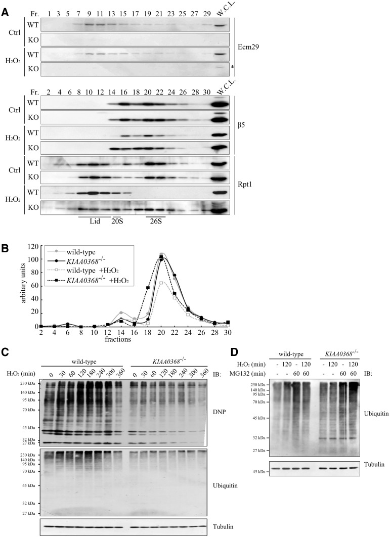
Fig. 5.
Analyses of proteasome complex upon oxidative stress. (A) Analysis of proteasome complexes under oxidative stress. Wild-type (WT) or KIAA0368-deficient (KO) MEF cells were treated with 100 μM H2O2 or H2O for 20 min. Five hundred micrograms of whole cell lysates were fractionated by glycerol density gradient (10–40% glycerol from 1 to 32 fractions) and immunoblotted with indicated antibodies. Asterisk indicates non-specific bands. W.C.L.; whole cell lysate. (B) Peptidase activities under oxidative stress. Odd fractions prepared in (A) were measured using Suc-LLVY-AMC. The vertical axis represent relative fluorescence normalized to peptidase activity of the first fraction. (C) Detection of oxidatively damaged proteins. Wild type and KIAA0368-deficient MEF cells were treated with 200 μM H2O2 for indicated times, then cell lysates were immunoblotted with indicated antibodies. (D) Accumulation of ubiquitylated proteins. Wild-type and KIAA0368-deficient MEF cells were treated with 200 μM H2O2 for 120 min in the presence or absence of 20 μM MG132 for 60 min, then the cell lysates were immunoblotted with indicated antibodies.

