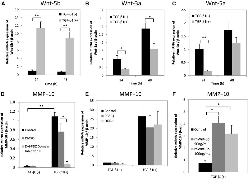
Fig. 4.
TGF-β1-induced MMP-10 expression seems to be mediated through non-canonical Wnt signalling possibly activated by Wnt-5b. (A–C) HSC-4 cells with (grey bars) or without (black bars) stimulation with 10 ng/ml TGF-β1 for 24 or 48 h in serum-free medium were analysed by qRT-PCR. The expression levels of (A) Wnt-5b, (B) Wnt-3a and (C) Wnt-5a were examined. Data are represented as the mean ± SD of three wells for each time point (*P < 0.05; **P < 0.01). HSC-4 cells with or without a 48-h stimulation using 10 ng/ml TGF-β1 were treated with (D) Dvl-PDZ Domain Inhibitor II (10 µM) or (E) DKK-1 (10 µg/ml) 60 min prior to TGF-β1 treatment. The expression levels of MMP-10 were examined by qRT-PCR. Data are represented as the mean ± SD of three wells for each time point (*P < 0.05; **P < 0.01). (F) HSC-4 cells were stimulated with 0, 50, or 100 ng/ml rhWnt-5b for 24 h in serum-free medium. The expression levels of MMP-10 were assessed by qRT-PCR. Data are represented as the mean ± SD of three wells for each time point (*P < 0.05).

