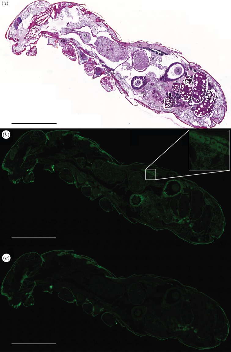
Figure 4.
Lateral panoramic views of a physogastric primary queen of Prorhinotermes canalifrons showing ChREBP expression in metabolic tissues. (a) Hemalin–eosin staining: ovaries (O); ovarioles (Ov); fat tissue (F); brain (B); nervous ganglions (GL); gut (G); and Malpighian tubules (MT). (b) ChREBP immunostaining (in green) using a commercial antibody generated against the human ChREBP peptide. (c) Control of ChREBP immunostaining. This view confirms that ChREBP-dependent fluorescence is not a result of the non-specific binding of secondary antibodies. Examples of non-specific signals are found in the external cuticle, the hindgut cuticle and as dense material (possibly urates) in dorsal and ventral clusters of cells localized in the parietal fat body below the cuticle (green autofluorescence). Results are representative of three independent experiments. Scale bars, 1 mm.

