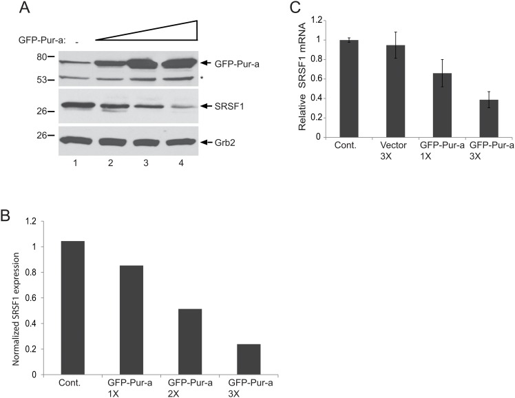
Fig 3. Dose dependent inhibition of SRSF1 by Pur-alpha in glial cells.
T98G cells were transfected with increasing concentrations of pEGFP-Pur-alpha plasmid (1X, 2X, and 3X) and whole cell protein extracts were prepared at 48hr post-transfection. Expression of SRSF1 and Pur-alpha were detected by Western blotting using anti-Pur-alpha and anti-SRSF1 antibodies. Grb2 was also probed in the same blots as an internal loading control. B. Signal intensities of SRSF1 expression in panel A were normalized to Grb2 levels and are shown as a bar graph. C. T98G cells were transfected with increasing concentrations of an expression plasmid encoding GFP-Pur-alpha. Total RNA was extracted and expression levels of SRSF1 mRNA transcripts were determined by Q-RT-PCR. mRNA levels of SRSF1 were normalized and are presented relative to change in relation to the control as a bar graph. All experiments were carried out in triplicate.

