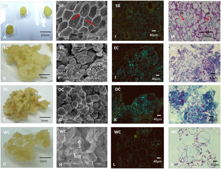
Fig 7. Morphology and histology of B. rotunda explant and callus.
A-D: morphology of samples; A: cross section of 1 cm x 1 cm shoot base tissue; B: friable pale yellowish callus; C: compact, dense and dry callus, D: spongy and wet callus; E-H: SEM images (100x magnification); E: regular-shaped and -sized cells with arrows showing the presence of starch; F: regular-shaped cells with fibrils; G: rounded, compact cells; H: elongated and irregular-shaped cells; I-L: morphology of each sample viewed under fluorescent microscopy with diphenylboric acid 2-aminoethylester (DPBA) stain (100x magnification); I: fluorescent yellowish-green lining of cell membrane, J: fluorescent greenish-blue spots observed with yellow lining of cell membrane; K: fluorescent greenish blue spots observed with yellow lining of cell membrane; L: yellowish lining of cell membrane; M-P: morphology of each sample viewed under light microscopy with Periodic acid Schiff (PAS) stain (100x magnification); E: organized and compact cells with presence of vascular bundles (VB) and purplish-red starch granules; F: presence of dark blue clusters indicates active cell division and purplish-red starch granules; G: presence of dark blue clusters indicates active cell division and red-purplish starch granules; H: irregular-shaped and -sized cells without starch granules. SB: shoot base; EC: embryogenic callus; DC: dry callus; WC: watery callus.

