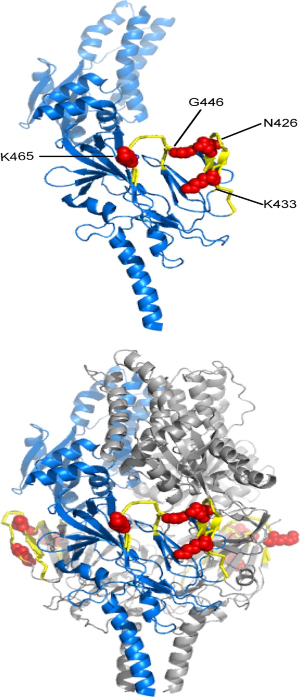
Fig 6. 2E1 binding epitope visualized on the RSV F prefusion structure.

The residues identified for 2E1 binding through shotgun mutagenesis are labeled and shown as red spheres and peptides identified through hydrogen/deuterium-exchange mass spectrometry are shown in yellow on the RSV F prefusion monomeric (top panel) and trimeric (bottom panel) structures [11].
