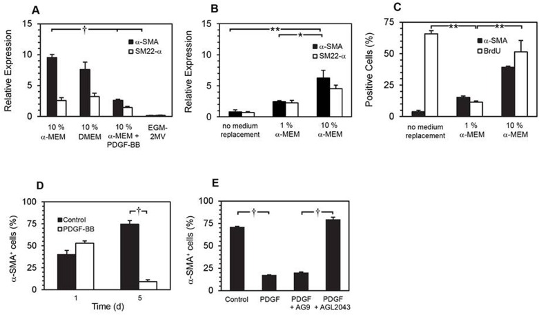
Fig 4. Effects of PDGF-BB on smooth muscle cell marker expression.
qRT-PCR analysis of α-SMA and SM22-α expression in BMCs cultured for 10 d in the specified media (A) († p < 0.001, Tukey-Kramer multiple comparisons test, n = 3). All BMCs were cultured in 10% α-MEM with additional PDGF-BB (50 ng/mL) for 8 d before replacing the media as indicated for 2 d. qRT-PCR analysis of α-SMA and SM22-α expression at 10 d, normalized against GAPDH (B). (* p < 0.05, ** p < 0.01, Bonferroni multiple comparisons test, n = 3). At 10 d, cells were immunolabelled for α-SMA and BrdU (** p < 0.01, Bonferroni multiple comparisons test, n = 3) (C). SMPCs were re-plated at day 10 in 10% α-MEM ± supplemental PDGF-BB (50 ng/mL) for 1 and 5 days, followed by immunofluorescence labelling of α-SMA (D) († p < 0.001, Bonferroni multiple comparisons test, n = 3). SMPC culture media was replaced with: control (no media change), media plus PDGF-BB (50 ng/mL), media plus PDGF-BB (50 ng/mL) and AG9 (10 μM), media plus PDGF-BB (50 ng/mL) and AGL2043 (10 μM), as indicated. Expression of α-SMA was detected by immunofluorescence microscopy after 5 d incubation (E) († p <0.001, Tukey-Kramer multiple comparisons test, n = 3).

