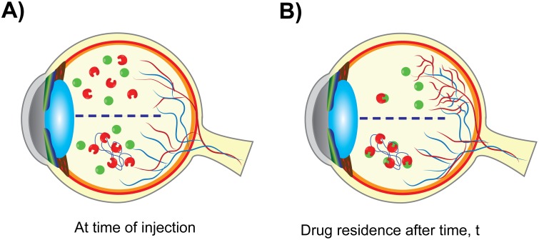
Fig 5. Schematic demonstrating the proposed mechanism of mvsFlt action.
A) sFlt (red, unconjugated) and mvsFlt (red conjugated blue chain of HyA) are injected into a diabetic retina where there is a high concentration of VEGF (green circles). B) After a given time, t, the majority of the sFlt has been cleared from the vitreous and VEGF is thus able to induce blood vessel growth. mvsFlt has a longer residence time in the vitreous and is able to bind and inhibit VEGF over much longer periods of time, leading to prolonged inhibition of retinal angiogenesis.

