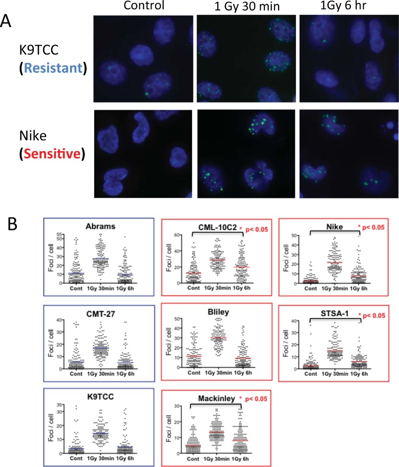
Fig 4. Phosphorylated-H2AX foci in G1 irradiated cells of radioresistant and radiosensitive groups of canine tumor cell lines.
Cells were synchronized in G1 using isoleucine deficient media and then irradiated with 1 Gy of gamma-rays. Following 30 min or 6 hr incubation time, cells were stained with γ-H2AX. (A) Examples of γ-H2AX foci (green) in nuclear DAPI (blue) staining in control, 1Gy followed by 30 min incubation and 1 Gy followed by 6 hr incubation in EdU negative cells. (B) Quantitative analysis of gamma-H2AX foci per cell. The pooled data from three independent experiments scoring 50 cells in each experiment are shown. The bar indicates mean. Statistical significances are shown for only control versus 6 h after 1 Gy (nonparametric Kruskal-Wallis test).

