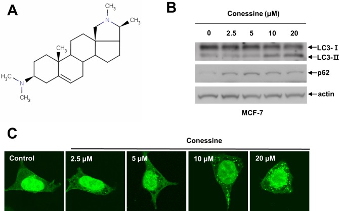
Fig 1. Conessine regulates autophagy.
(A) Chemical structure of conessine. (B) Conessine treatment increased the level of LC3-II and p62. MCF-7 cells were incubated with the indicated concentrations of conessine (0, 2.5, 5, 10, and 20 μM) for 24 h, and the cell lysates were subjected to Western blotting with the indicated antibody. (C) Conessine treatment induced autophagosome formation in HEK293 cells. HEK293 cells stably expressing GFP-LC3 were incubated with the indicated concentration of conessine for 24 h, and the cells were analyzed with confocal microscopy.

