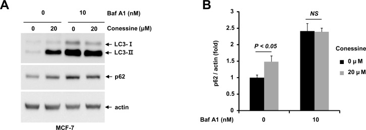
Fig 3. Conessine interferes with autophagic flux.
(A) Conessine treatment suppressed with autophagic flux. MCF-7 cells were treated with either mock or conessine (10 μM) in the presence or absence of Bafilomycin A1 (10 nM). (B) The levels of p62 were analyzed. Autophagic flux experiments were performed in triplicate, and the mean and standard deviations are shown in the graph.

