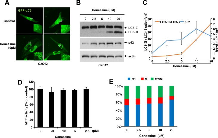
Fig 4. Conessine treatment induces autophagosome formation in C2C12 cells.
(A) C2C12 cells were transfected with a plasmid encoding GFP-LC3 and cells were treated with either mock or conessine (10 μM). (B) Conessine treatment increased the level of LC3-II and p62. C2C12 cells were incubated with the indicated concentration of conessine for 24 h, and the cell lysates were subjected to Western blotting with the indicated antibody. (C) The levels of LC3-II/LC3-I and p62 were analyzed. The experiments were performed in triplicate, and the means and standard deviations are shown in the graph. (D) Conessine less than 10 μM did not affect proliferation of C2C12 cells. C2C12 cells were treated with the indicated concentration of conessine, and cell viability were measured by MTT assay (E) Conessine treated cells were analyzed with flow cytometry. The percentage of cells at G1, S and G2/M was measured, and is shown in the graph.

