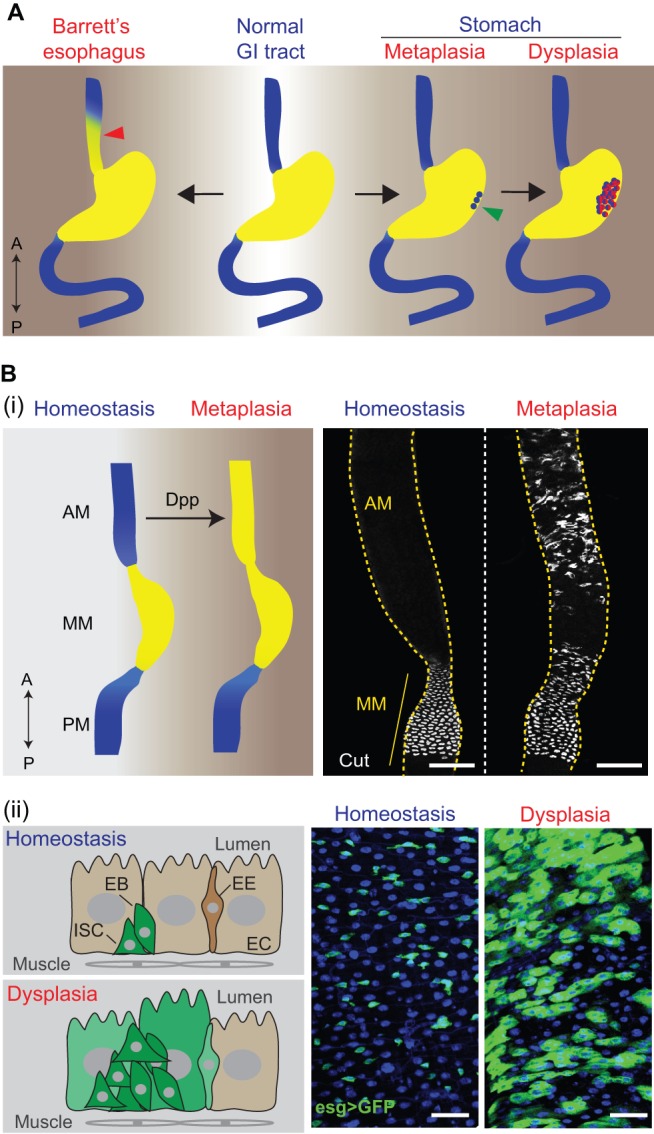
Fig. 3.

Metaplasia and dysplasia in the human and Drosophila gastrointestinal (GI) tract. (A) GI pathologies in humans (‘A’, anterior, top; ‘P’, posterior, bottom). Barrett's esophagus (left) is a metaplastic lesion, in which the distal esophageal epithelium (see red arrowhead) is replaced by a stomach- or intestine-like columnar epithelium. Gastric metaplasia (right) is another common metaplastic disease in the human GI tract, in which intestinal epithelial cells (see green arrowhead) are found in the stomach. Gastric metaplasia can develop further into dysplasia (far right, depicted in red), characterized by atypical changes of the epithelial structure due to aberrant cell proliferation and differentiation. (B) GI pathologies in Drosophila. (i) Metaplasia in the Drosophila anterior midgut (AM) is characterized by ectopic generation of copper cells (CCs), normally restricted to the middle midgut (MM) in homeostatic conditions, in the AM. The schematic on the left summarizes the replacement of AM cells by MM cells (depicted in yellow) during metaplasia. PM, posterior midgut. The images on the right were obtained by immunostaining (using an antibody specific for Cut, a marker for CCs) and confocal microscopy of the midgut region in flies. Dpp overexpression (right-hand panel) generates ectopic acid-producing CCs, indicative of metaplasia (see main text for further details). Scale bars: 100 μm. Microscopy images adapted from Li et al. (2013a). (ii) Dysplasia in the Drosophila midgut is characterized by stem cell over-proliferation and abnormal differentiation. The schematic on the left summarizes changes of the epithelial structure during dysplasia. The images on the right were obtained from confocal microscopy of the posterior midgut (blue, DAPI; green, GFP). The stem/progenitor cell marker esg>GFP (esg::Gal4, UAS::GFP) is only expressed in intestinal stem cells (ISCs) and enteroblasts (EBs) during homeostasis, whereas, in the dysplastic condition, esg>GFP begins to be expressed in differentiated cells, including enterocyte (EC)-like cells and enteroendocrine cells (EEs). Scale bars: 25 μm.
