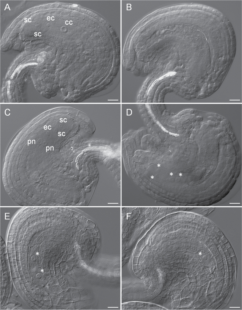
Fig. 4.
Female gametophyte development in WT and ubc22-1. Ovules from flowers just before opening were prepared and observed under a microscope with DIC. (A) A typical WT embryo sac (FG7 stage) showing one central cell (cc), one egg cell (ec), and two synergid cells (sc). (B–F) Abnormal embryo sacs in ubc22-1 plants. (B) Mutant embryo sac without any nucleus. The embryo sac was smaller and narrower than the WT embryo sac. (C) Mutant embryo sac at the FG5 stage showing two polar nuclei (pn), two synergid cells, and one egg cell. (D) Mutant embryo sac at the FG4 stage, with four nuclei (indicated by asterisks). (E) Mutant embryo sac at the FG3 stage, with two nuclei and a larger vacuole between them. (F) Mutant embryo sac with only one nucleus. Scale bars, 10 μm.

