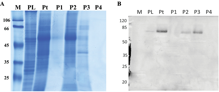
Fig. 2.
SDS-PAGE (a) and immuno blotting (b) of proteins extracted from leaves of C. plantagineum. Note: M, protein size markers (given in kDa); PL, proteins not soluble in extraction buffer; Pt, total proteins extracted by the extraction buffer; P1, proteins precipitated by 0–25% (w/v) (NH4)2SO4; P2, proteins precipitated by 25–50% (w/v) (NH4)2SO4; P3, proteins precipitated by 50–70% (w/v) (NH4)2SO4; P4, proteins remaining in extraction buffer after precipitation by 50–70% (w/v) (NH4)2SO4; all pellets were dissolved in buffer A of the same volume as used in the extraction. Samples were loaded with SDS buffer on the gel.

