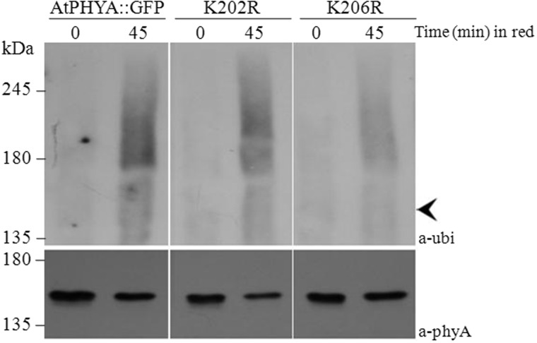
Fig. 3.
Western blot analysis of phytochrome-ubiquitin conjugates of wild type, K202R and K206R phyA proteins. Proteins were extracted from 3-day-old etiolated seedlings exposed to continuous red light (12 µmole m−2 s−1) and immunoprecipitated using anti-GFP polyclonal antibody. Immunoprecipitated samples were separated on 6% SDS-PAGE gels and probed with anti-Ubi or anti-phyA antibodies. Arrowhead indicates unubiquitinated AtPhyA::GFP (∼150 kDa).

