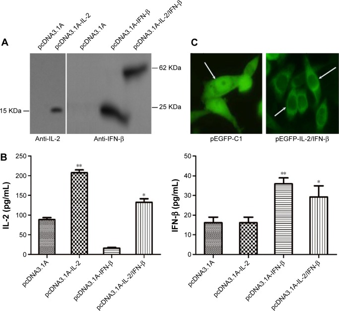
Figure 1.
The expression of IL-2 and IFN-β fusion protein in Lovo cells.
Notes: (A) The expression of pcDNA3.1A-IL-2, pcDNA3.1A-IFN-β, and pcDNA3.1A-IL-2/IFN-β was detected by Western blot analysis in Lovo cells using specific antibodies. (B) ELISA was performed to detect the IL-2/IFN-β fusion protein levels in supernatants of medium. Column: mean (n=3); bar: SD. *P<0.05 vs pcDNA3.1A; **P<0.01 vs pcDNA3.1A. (C) Subcellular distribution of IL-2/IFN-β-GFP and GFP in Lovo cells. Arrows represent the location of GFP. The control GFP was expressed in both the cytoplasm and nucleus, while IL-2/IFN-β-GFP fusion protein was mainly located in the cytoplasm of Lovo cells.
Abbreviations: IL-2, interleukin-2; IFN-β, interferon-β; ELISA, enzyme-linked immunosorbent assay; SD, standard deviation.

