Figure 6.
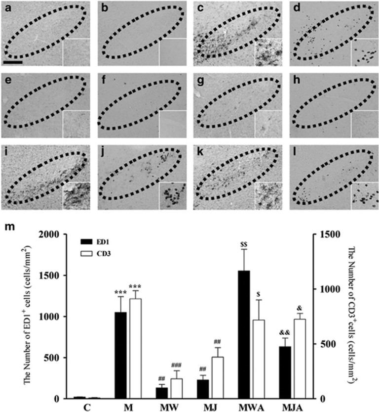
The CB2 receptor inhibits MPTP-induced infiltration of peripheral immune cells in the SN in vivo. The SN tissues obtained from the same animals used in Figure 5 were immunostained with an ED1 antibody to label phagocytotic macrophages and microglia (a, c, e, g, i, and k) and with a CD3 antibody to label T cells (b, d, f, h, j, and l). Animals that received PBS as a control (a, b); MPTP (c, d); MPTP and WIN55,212-2 (e, f); MPTP and JWH-133 (g, h); MPTP, WIN55,212-2 and AM630 (i, j); or MPTP, JWH-133 and AM630 (k, l) were killed 3 days after the last MPTP injection. Insets show higher magnifications of a–l. Dotted lines indicate the SNpc. Scale bars: a–l, 200 μm. (m) The number of CD3+ (white bars) or ED1+ (black bars) cells in the SN were counted. Four to five animals were used for each experimental group. C, control; M, MPTP; MW, MPTP and WIN55,212-2; MJ, MPTP and JWH-133; MWA, MPTP and WIN55,212-2 and AM630; MJA, MPTP and JWH-133 and AM630. ***P<0.01 significantly different from controls; ##P<0.01 and ###P<0.001 significantly different from MPTP only; $P<0.05 and $$P<0.01 significantly different from MW; &P<0.05 and &&P<0.01 significantly different from MJ (ANOVA and Student–Neuman–Keuls analysis).

