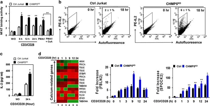
Figure 5.
CHMP5-knockdown Jurkat cells enhance the activation of NFAT in response to TCR. (a) Ctrl and CHMP5KD Jurkat cells were stimulated with anti-CD3/anti-CD28 for different times or 10 ng ml−1 PMA and 1.5 μM ionomycin in the presence or absence of 100 nM CsA, as a positive control. Nuclear extracts were prepared and analyzed for DNA-binding activities of NFAT. All error bars represent ±s.d. of the mean from triplicate samples. *P<0.01 and **P<0.05. (b) Ctrl and CHMP5KD Jurkat cells were treated with or without anti-CD3/anti-CD28 at different times, as indicated. The levels of intracellular IL-2 were measured by flow cytometry. The results represent ±s.d. of the mean from triplicate samples. (c) Ctrl and CHMP5KD Jurkat cells were stimulated with or without anti-CD3/anti-CD28 for 24 h, and the amount of IL-2 in the supernatant was measured by ELISA. All error bars represent ±s.d. of the mean from triplicate samples. *P<0.05. (d) RNA was extracted from Ctrl and CHMP5KD Jurkat cells treated with anti-CD3/anti-CD28 for 1, 3, 9, 12 and 24 h, followed by analysis of calcium-related gene expression in Ctrl and CHMP5KD Jurkat cells (CHMP5KD versus Ctrl Jurkat cells). Lane 1, Unstimulated cells; Lane 2, TCR stimulation for 1 h; Lane 3, TCR stimulation for 3 h; Lane 4, TCR stimulation for 9 h; Lane 5, TCR stimulation for 12 h; Lane 6, TCR stimulation for 24 h. RNA was extracted from cells, and then q-RT-PCR analysis was performed with specific primers targeted to FBLN2 and SPOCK2, respectively. Data represent the average of data from two independent experiments, each conducted in triplicate. Error bars represented the mean±s.d. of six samples. *P<0.01 and **P<0.05.

