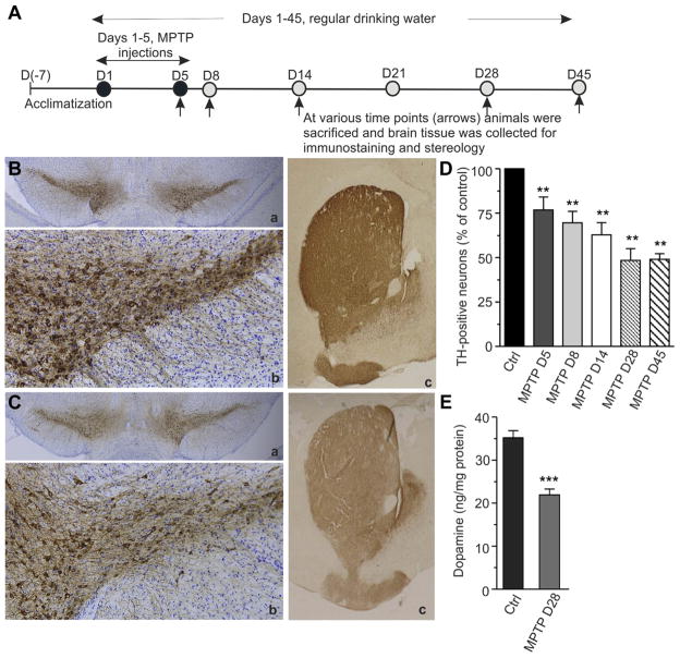
Fig. 2.
Progressive loss of TH-positive neurons in subchronic MPTP model. (A) Schematic outline of the experimental design. Mice were acclimatized to the new environment for 7 days before the start of the experiment. Animals were randomly divided into experimental groups and given 5 daily injections of MPTP (25 mg/kg/injection). Control mice were injected with saline. On days 5, 8, 14, 28, and 45 after the MPTP injections (arrows), mice were sacrificed and brain tissue was collected for immunohistochemistry (B and C), stereology (D), and biochemical (E) analyses. Other details as described in Section 2. (B and C) Brains were fixed and immunostained with rabbit polyclonal anti-tyrosine hydroxylase antibody (brown) and counterstained with cresyl violet (blue) for anatomic reference. Images were captured on an Olympus microscope equipped with Microcast 3CCD 1080p HD color camera. (B) Representative photomicrographs of midbrain sections from control mouse showing TH-immunoreactivity. In B (a) coronal section of mouse midbrain across both hemispheres showing intense brown staining of SNpc region; B (b) shows dense network of TH-immunopositive cell bodies and fibers in the SNpc; B (c) shows strong TH-immunoreactive dopamine terminals in the striatum. (C) Representative photomicrographs of midbrain sections from MPTP-injected mouse showing reduced TH-immunoreactivity (pale brown) on D28. In C (a) reduced TH staining of SNpc (pale brown); C (b) decreased density of TH-immunopositive cell bodies and fibers in SNpc; C (c) reduction in TH-immunoreactive dopamine terminals in the striatum. Magnifications: B (a), B (c), C (a), and C (c) = 2.5×; B (b) and C (b) = 10×. (D) Progression of dopaminergic neurodegeneration over a period of 45 days. TH-positive cells were counted using an unbiased stereology method and cell survival was plotted as percentage of control. Results are shown as mean ± SEM (n = 5–6 per group from a single experiment). Statistically significant differences between control and MPTP-injected groups are shown as ** p ≤ 0.01, for all groups. (E) Reduction in the striatal dopamine level during the course of neurodegeneration. Brain tissue was snap-frozen in liquid nitrogen and dopamine levels were analyzed by HPLC. Results are shown as mean ± SEM (n = 8–10 per group from 5 separate experiments). Statistically significant difference between control and MPTP-injected group is indicated as *** p ≤ 0.001. Abbreviations: MPTP, 1-methyl-4-phenyl-1, 2, 3, 6-tetrahydropyridine; SNpc, substantia nigra pars compacta; TH, tyrosine hydroxylase.

