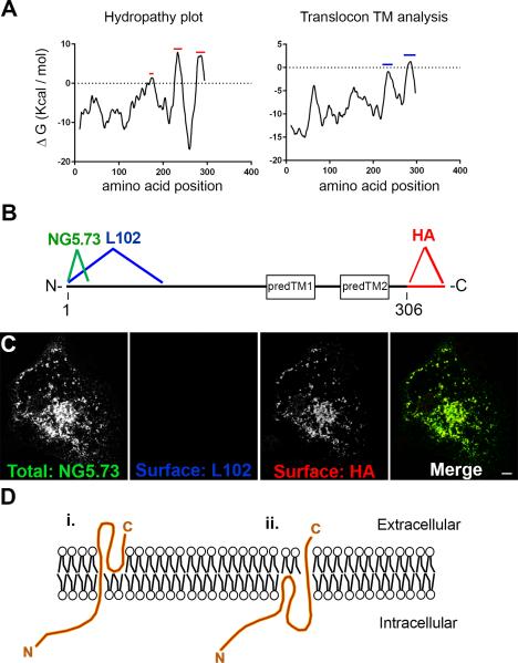
Figure 2. SynDIG4 topology.
A: Hydropathy plot and translocon TM analysis show three predicted hydrophobic domains (red bars) but only two predicted transmembrane domains (blue bars).
B: Schematic of SynDIG4-HA protein and two predicted transmembrane domains. The antigen sites for NG5.73 and L102 are located near the N-terminus, and the HA tag is on the C-terminus. Regions predTM1 and predTM2 represent the hydrophobic domains that are predicted to be transmembrane.
C: Representative image of COS cells transfected with SynDIG4-HA and surface labeled with L102 (blue) and HA (red) antibodies. NG5.73 (green) was used to stain for total SynDIG4 after fixation and permeabilization. Scale bar = 10 μm.
D: Possible models for SynDIG4 topology, with the C-terminus extracellular, and the N-terminus intracellular. The loop region between the two predicted transmembrane domains could either be extracellular (i) or intracellular. Models are not to scale.

