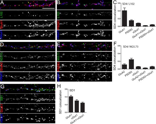
Figure 9. SynDIG4 is enriched at extrasynaptic AMPARs.
A, B, D, E, G: Representative dendrite stretches of dissociated primary cortical neurons (10 DIV) stained with antibodies against SynDIG4 (SD4) or SynDIG1 (SD1) and other synaptic markers. A, B: SD4/ L102/45, D, E: SD4/NG5-73, and G: SD1. GluA1, GluA2, and PSD93 were used as post-synaptic markers, with vGlut1 as a presynaptic marker. Synapses were defined as overlapping pre- and post-synaptic markers.
C, F, H: Quantification of (A-B, D-E, and G), respectively, represented as the fraction of SynDIG1 or SynDIG4 colocalized with the indicated synaptic marker (n = 10 for each staining set). Scale bar = 10 μm.

