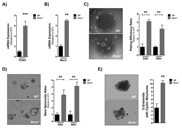
Figure 3. Bves−/− enteroids exhibit increased stemness ex vivo.
Small intestinal crypts were isolated from WT or Bves−/− mice and embedded in Matrigel. (A) qRT-PCR analysis revealed increases in (A) PCNA (***P<0.001, n=6) and (B) Muc2 (**P<0.01, n=6) mRNA levels in Bves−/− enteroids compared to WT. Enteroid stem cell properties determined based on (C) plating efficiency ratio, as measured by percentage of surviving enteroids 24 and 48 hours post-plating compared to total crypts plated (24 hours, **P<0.01; 48 hours, **P<0.01; n=6); (D) ratio of stem spheroid proportions counted 24 and 48 hours post-plating (24 hours, **P<0.01; 48 hours, **P<0.01; n=6) and (E) percentage of enteroids maintaining peripheral cystic structures 5 days after passaging (3.8 ± 1.0% vs. 10.2 ± 0.6%, **P<0.01, n=6). Images were captured at 40× magnification (3C) or 100× magnification (3D, 3E).

