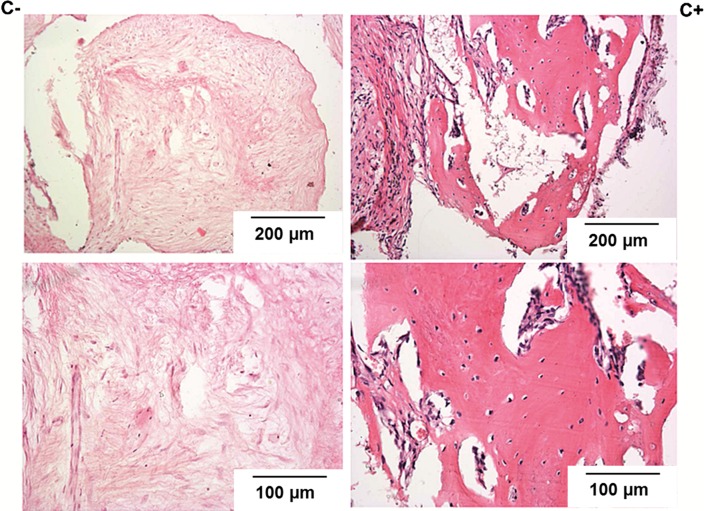
Figure 7.
Histological pictures of the explants harvested 12 weeks after implantation (HE staining)—higher magnification at the bottom line. On the left side—pictures from the group where the naked, i.e. not-seeded with cells, scaffolds were implanted (C−). On the right side—pictures from the group where the implanted scaffolds were seeded with cells prior to implantation (C+). Only in calcites seeded with HBDCs before implantation bone tissue was found within the implants.

