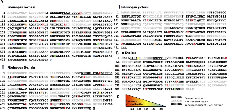
Figure 5.
Mass spectrometry analysis of peptides formed by RgpB and PPAD from fibrinogen and α-enolase. Substrates were incubated o/n at 37°C with 10:1 concentration of RgpB and tPPADWT in ddH2O. Peptides formed were identified by liquid chromatography-mass spectrometry (LC-MS), and occurrence of citrullinated variants quantified. (A) Peptides formed from fibrinogen α-chain, β-chain and γ-chain (panels i, ii, and iii, respectively). (B) Peptides formed from α-enolase. (C) Key: Sequence in black denotes region covered in LC-MS result, grey depicts non-covered regions. Underlined sequences denote known immunodominant B cell epitopes. Arginine residues occurring frequently citrullinated are represented by red/orange R, occasionally citrullinated by yellow/green R, and those never detected citrullinated by blue R.

