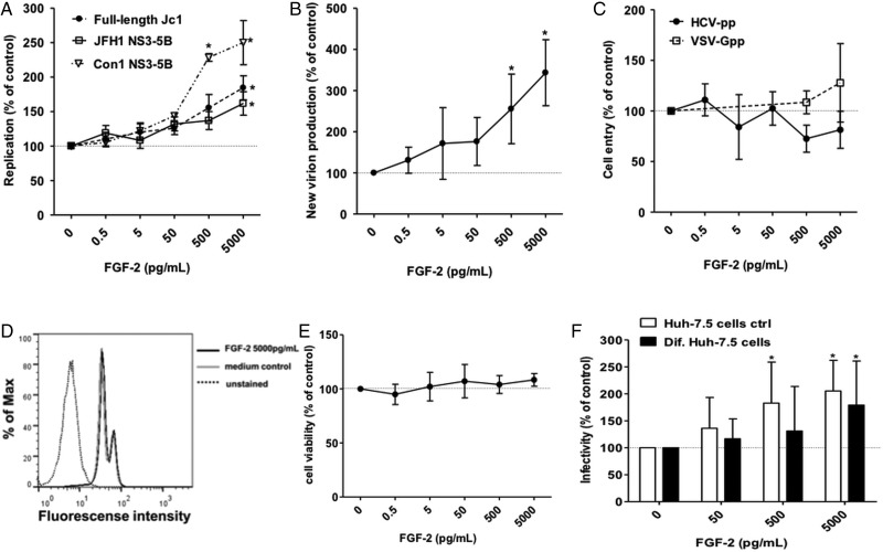
Figure 3.
Fibroblast growth factor-2 (FGF-2) enhances HCVcc replication and new particle production in a concentration dependent manner. (A) Huh-7.5 cells were electroporated with the indicated Fluc-reporter subgenomic or full-length HCV genomes RNA. Five hours later media supplemented with the indicated concentration of FGF-2 was added. Cells were lysed and luciferase measured after 48 h of incubation. (B) Supernatants from the full-length Jc1 experiment represented in panel A were collected 48 h after electroporation, filtered and used to infect naïve Huh-7.5 cells. After 6 h media was exchanged and the cells were incubated without FGF-2 for 48 h before lysis and quantification of luciferase activity. (C) Huh-7.5 cells were transduced with HCVpp (clone H77) and VSV-Gpp in the presence of FGF-2 at the indicated concentrations. (D) Huh-7.5 cells were exposed to FGF-2 for 48 h. Then cells were fixed and effect of FGF-2 on cell proliferation was assessed by reading incorporation of 5-ethynyl-2′-deoxyuridine into cellular DNA by flow cytometry. The analysed image is of the highest tested concentration of FGF-2. Medium without FGF-2 was used as control in parallel. (E) Huh-7.5 cells were exposed to FGF-2 for 48 h. Then the effect of FGF-2 on cell viability was assessed by detecting release of intracellular proteases using the CytoTox-Glo Cytotoxicity Assay. (F) Differentiated non-dividing Huh-7.5 cells were infected with Fluc-Jc1 HCVcc. After 6 h cells were washed and treated with FGF-2 at the indicated concentrations. After 48 h of incubation cells were lysed and luciferase expression was quantified. Regular dividing Huh-7.5 cells were used as control in parallel. In each panel, dots or bars represent the mean±SD of minimum n=6 replicates from three independent experiments with the mean value obtained in the absence of FGF-2 set to 100%.

