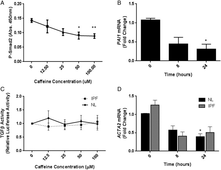
Figure 1.
(A) Immortalised human bronchial epithelial cells (iHBECs) were stimulated with increasing concentrations of caffeine for 4 hours and PSmad2 levels measured. Figure shows mean data±SEM from three independent experiments. (B) iHBECs were stimulated with 50 µM caffeine and PAI1 mRNA levels measured. Data are expressed as mean fold change over control (0 h)±SEM from three independent experiments. (C) Non-fibrotic control (NL) and idiopathic pulmonary fibrosis (IPF) fibroblasts were stimulated with increasing concentrations of caffeine and TGFβ activation assessed by TMLC reporter assay. Figure shows mean data±SEM from n=3 NL and n=3 IPF donors. (D) NL and IPF fibroblasts were stimulated with 50 µM caffeine and ACTA2 mRNA levels measured. Data are expressed as mean fold change over control (0 h for NL or IPF, respectively)±SEM. Figure shows mean data from n=3 NL and n=3 IPF donors. *p<0.05 **p<0.01.

