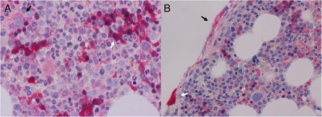
Figure 1.
Normal bone marrow stained for transforming growth factor α. (A) Erythroid cells are moderately to strongly positive (white arrow) and megakaryocytes are weakly positive (black arrow). Magnification ×600. (B) Intensely positive osteoclast adjacent to trabecular bone (white arrow) with moderate intensity staining in multiple osteoblasts (black arrow). Magnification ×400.

