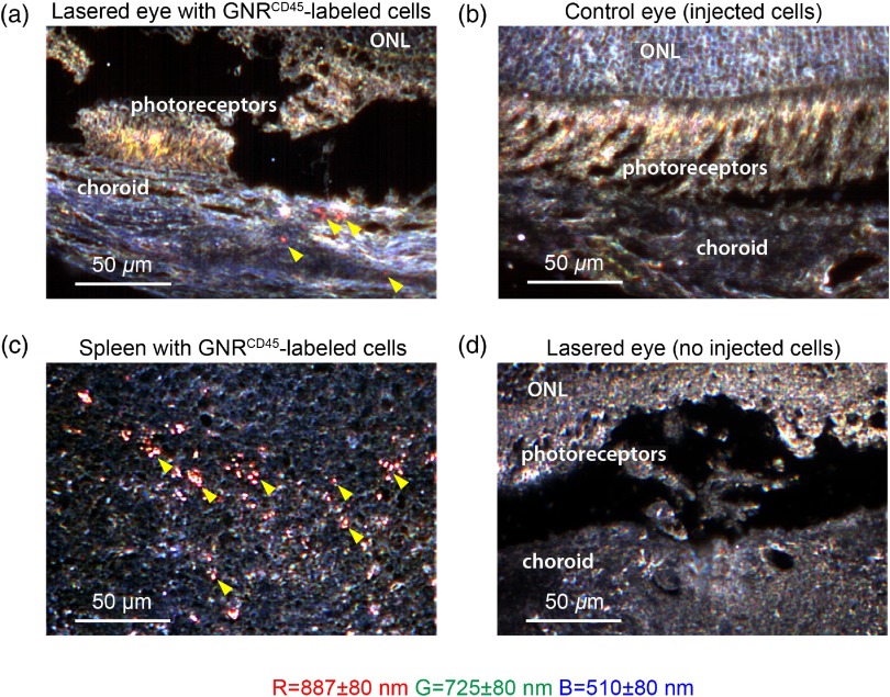
Fig. 6.
Hyperspectral images of GNR-labeled leukocytes inside lasered eyes of mice. (a) Hyperspectral image of -thick unstained section of lasered eye and (b) the corresponding nonlasered control eye of nu/nu mice that received -labeled cells. The choroid, photoreceptor, and outer nuclear layer (ONL) regions of the retina have been marked inside the image. Red GNR-labeled cells are indicated by yellow arrowheads. (c) Hyperspectral image of -thick unstained section of spleen of mice injected with GNR-labeled cells. Red GNR-labeled cells indicated by yellow arrowheads. (d) Hyperspectral image of -thick unstained section of laser-treated eye of control mice without injected GNR-labeled cells. Center of bands are , , ; each band is Gaussian with an FWHM of 80 nm. .

