Fig. 10.

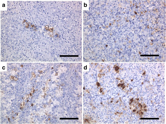
Immunohistochemical stainings of the original tumor (a, c) and heterotransplanted tumor (b, d) with CA 125 (a, b) and CA 19–9 (c, d). Cancer cells were positive for CA 125 and CA 19–9 (a, b, c, d; bar = 100 μm)

Immunohistochemical stainings of the original tumor (a, c) and heterotransplanted tumor (b, d) with CA 125 (a, b) and CA 19–9 (c, d). Cancer cells were positive for CA 125 and CA 19–9 (a, b, c, d; bar = 100 μm)