
Fig. 14.
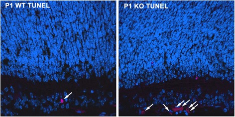
Increased apoptosis in the ganglion cell layer of AP-2δ−/− P1 retina. Apoptotic cells in P1 retinal tissue sections from AP-2δ+/+ (WT) and AP-2δ−/− (KO) mice were stained using the In Situ Cell Death Detection kit, TMR red (Roche). Photographs were taken using a Zeiss LSM510 confocal microscope. Arrows point to apoptotic cells