
Fig. 4.
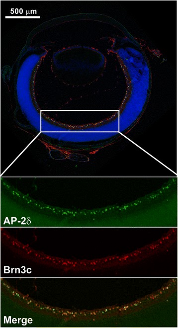
Co-expression of AP-2δ and Brn3c in retinal ganglion cells. P1 retinal tissue sections were immunostained with anti-AP-2δ and anti-Brn3c antibodies followed by secondary antibodies conjugated to Alexa 488 and Alexa 555, respectively. Sections were counterstained with DAPI to label the nuclei. Images were automatically assembled from multiple scans (4 × 4 scan; 4096 × 4096 pixels) using the Tile-scan function of the LSM program. The area outlined by the rectangle is magnified to show AP-2δ-positive cells (green), Brn3c-positive cells (red) and merged AP-2δ and Brn3c