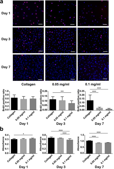
Fig. 5.

Proliferation of cardiac fibroblasts cultured on collagen and CNH-Col substrates on days 1, 3, and 7. a BrdU staining, where the red immunofluorescence represents proliferative cardiac fibroblasts (bars = 60 um). The histogram shows that there are significant differences in the BrdU-positive rate between the collagen and CNH-Col substrates at different stages, especially for the 0.1 mg/ml CNH-Col group. b The CCK-8 assay shows that there are significant differences in the absorbance of cardiac fibroblasts cultured on collagen and CNH-Col substrates at different stages. Data are means ± SDs, n = 3
