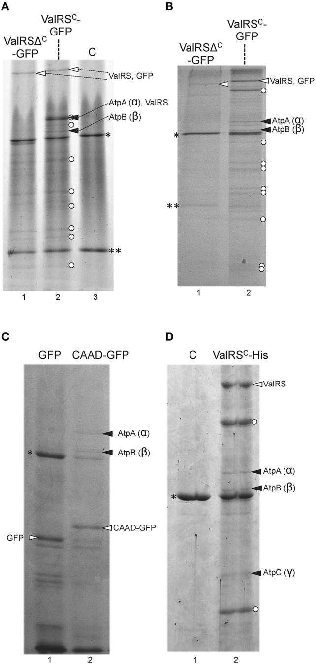
Figure 5.

Co-purification of Anabaena ValRSC with subunits of the ATP synthase. (A) Whole cell extracts from Anabaena strains expressing the fusion protein indicated at the top of the panel were purified using anti-GFP antibodies and resolved by SDS-PAGE gel. Control lane labeled as “C” contains purification fractions of Anabaena cells not expressing any fusion protein. White arrowheads point to the corresponding fusion protein. Black arrowhead point to subunits of ATP synthase. White dots indicate proteolytic fragments of the GFP fusion proteins. Asterisks indicate the position of RubisCO, double asterisks indicate the position of the light chain of inmunoglobulins (B) and (C) Membrane fractions of Anabaena strains expressing the fusion protein indicated at the top of the panel were purified using anti-GFP antibodies and resolved by SDS-PAGE gel. Other details are like in (A). (D) Membrane fractions of Anabaena strains expressing the fusion protein indicated at the top of the panel were purified using anti-hexahistidine antibodies and resolved by SDS-PAGE gel.
