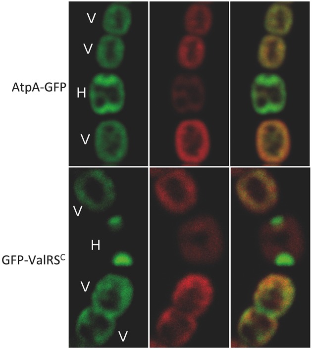
Figure 8.

Subcellular localization of AtpA. Pictures correspond to confocal microscopy images of Anabaena cells expressing the protein indicated at the left margin. Left panels show the green fluorescence of GFP, central panels the red fluorescence of photosynthetic pigments and the right panel the merged picture of the former ones. V, vegetative cells; H, heterocyst.
