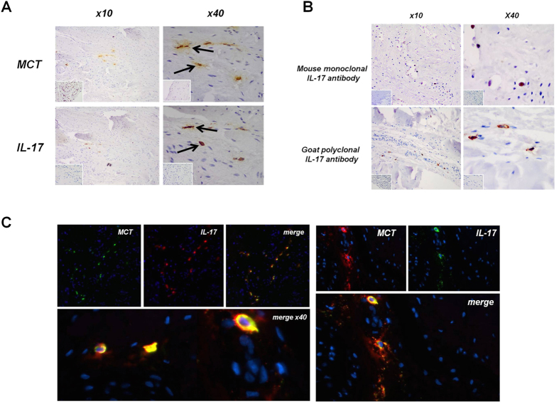
Figure 2. IL-17A localisation in tendon samples.
(A) Early human tendinopathy sample (matched subscapularis tendon biopsy) stained for (A) Mast cell tryptase (MCT) and IL-17A, Isotype IgG in bottom left corner using goat polyclonal IL-17A antibody. (B) Early tendinopathy sample stained for IL-17A using mouse monoclonal antibody and goat polyclonal antibody. Isotype IgG in bottom left corner. (C) Double imunofluorescence staining using goat polyclonal IL-17A antibody showing IL-17A, Mast Cell Tryptase and double staining co-localization in early tendinopathy sample. (x100).

