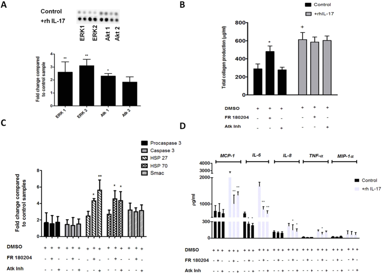
Figure 5. IL-17A induced phosphorylation of MAPK in cultured tenocytes and matrix response to ERK and Atk inhibition.
(A) Whole-cell lysates from tenocytes were examined for the expression of phosphorylated ERK1, ERK2, Atk1 and Atk2 24 hours after exposure to 100 ng rhIL-17A The fold change of MAPK’s was determined by densitometry and normalised to the control sample on the array. Data are shown as the mean fold change ± SD of duplicate samples and are representative of experiments using three individual donors of normal hamstring tendon explant tissue. *p < 0.05, **p < 0.01 compared to control samples (ANOVA). (B) Cells were preincubated for 24 h with specific inhibitors for ERK (FR 180204)(10 μM), or Atk(5 μM) which also were included in the assay media. Total collagen production was assessed using the Sircol assay *p < 0.05, **p < 0.01 (Students t-test). (C) The levels of apoptotic proteins induced following incubation with ERK and Atk inhibitors were determined (apoptotic proteome profiler) by densitometry and normalised to the relevant control sample on the array. Data are shown as the mean fold change ± SD of duplicate samples and are representative of experiments using five individual donors of normal hamstring tendon explant tissue *p < 0.05, **p < 0.01 (ANOVA). (D) Cells were preincubated for 24 h with 100 ng recombinant IL-17A and specific inhibitors for ERK (FR 180204) and Atk, which also were included assay media. Data shows levels of MCP-1, IL-8, IL-6, TNF-α and MIP-1α in supernatants removed from culture analysed using Luminex. Data shown are the mean ± SD of triplicate samples and are representative of five normal hamstring tendon explant experiments *p < 0.05, **p < 0.01 (Students t-test).

