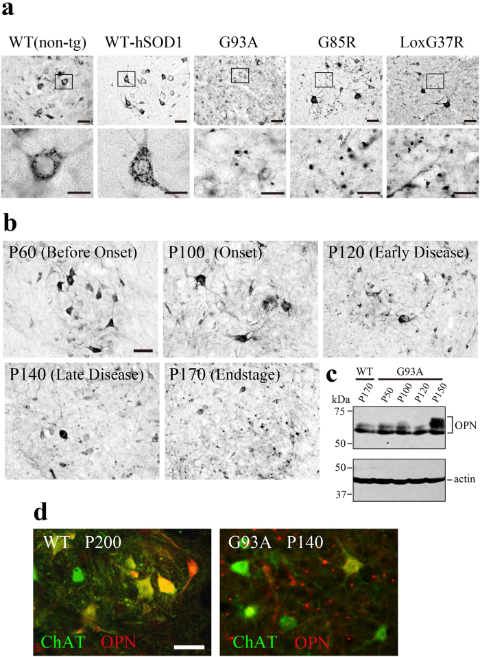
Figure 3. Accumulation of extracellular OPN-immunopositive granules in the spinal cords of ALS model mice during disease progression.
(a) OPN immunostaining in lumbar spinal cord sections from SOD1G93A (G93A), SOD1G85R (G85R) and LoxSOD1G37R (LoxG37R) mutant mice at their respective end stages, or from non-transgenic control mice (WT) at P200 or human wild-type SOD1-transgenic mice (WT-hSOD1) at P180. OPN signals were detected within MN cell bodies in the WT and WT-hSOD1 mice. Extracellular, granule-like deposits were scattered outside the perikarya in the ALS mutant lines. Boxed areas in the upper panels are enlarged in the lower panels. (b) Changes in the distribution of OPN-immunopositive signals during the disease course in SOD1G93A mice. OPN-positive extracellular granules became detectable around the time of disease onset (P100) and increased in number as the disease progressed (P120–P170). (c) Western blot analysis of spinal cord lysates from non-transgenic control (WT) and SOD1G93A mice (G93A) prepared at the indicated times. Representative blot out of three experiments. Full-length gels are shown in Supplementary Fig. 10a. (d) Double-immunofluorescent labeling of lumbar spinal cord sections from WT (P200) and SOD1G93A (P140) mice shows the appearance of OPN-positive extracellular granules in the vicinity of ChAT-positive motor neurons in SOD1G93A mice. Scale bar, 50 μm (a upper panels, b, d); 20 μm (a lower panels). See also Supplemental Fig. 3.

