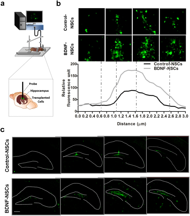
Figure 2. Engrafted NSCs Survive and Migrate in the AD Hippocampus.
(a) Schematic drawing represents how to use the fluorescence endoscopic confocal microscopy system on living AD mice. (b) Top, representative images of living engrafted GFP-expressing cell distributed from top to bottom in the hippocampus of Tg+Control-NSCs and Tg+BDNF-NSCs mice at 4 weeks after transplantation. Each dashed line indicates the vertical extensive position of the acquired view. Quantification of the relative fluorescence units within the vertical extensive path shows that more GFP signals are detected in Tg+BDNF-NSCs mice. (c) IF staining of engrafted GFP-expressing cells 4 weeks after transplantation shows that engrafted GFP-expressing cells are able to survive and migrate in the hippocampus of AD mice. Scale bar: 200 μm.

