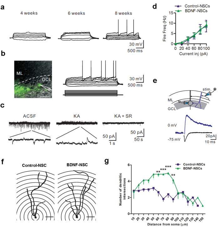
Figure 6. Engrafted NSC-derived Neurons Display the Electrophysiological Properties of Mature Neurons and Exhibit Neurite Complexity.
(a) Representative voltage responses of engrafted cells at 4, 6, and 8 weeks after transplantation. Data represent by BDNF-NSC. (b) Left, a representative recording of an engrafted cell. Scale bar: 20 μm. Right top, voltage responses of the recorded cell; right bottom, 2 s current protocol, 10 pA steps. Data represent by BDNF-NSC. (c) Spontaneous synaptic currents recorded from the recorded cell in (b) holding at −55 mV. The enlarged trace indicates the spontaneous EPSCs and IPSCs. The spontaneous events were blocked by the addition of AMPA/NMDA (KA) and GABAergic receptor blockers (SR). (d) Mean firing frequency of Control-NSC- and BDNF-NSC-derived neurons 8 weeks after transplantation (Control-NSCs, n = 16 from 5 mice; BDNF-NSCs, n = 19 from 7 mice). (e) Top, experimental diagram. Stimulation electrode was placed in the molecular layer at a distance of 100 μm from the recorded cell. Bottom, the evoked AMPAR-/NMDAR- (black) or GABAR-mediated (blue) currents of the recorded cell in (b) (average from 10 sweeps). (f) Representative reconstructed cell of Control-NSC-derived and BDNF-NSC-derived neuron with the given radius. Scale bar: 20 μm. (g) Sholl analysis for both kinds of cell-derived neurons. (Control-NSCs, n = 6 from 3 mice; BDNF-NSCs, n = 7 from 3 mice; results presented as the mean ± SEM). Statistical analyses by two-way ANOVAs with Bonferroni post-tests.

