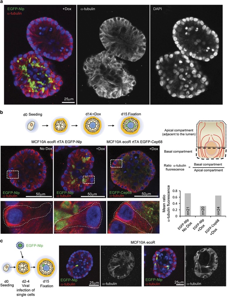
Figure 3.
Nlp CRBs alter the organization of MTs in polarized epithelia of mammospheres. (a) Impact of Nlp CRBs on MT organization in mammospheres. Micrographs show juxtaposition of two mammospheres, only one of which expresses Nlp in response to Dox. Left panel: merge of pseudo-colors illustrating EGFP-Nlp in green, α-tubulin in red and DNA in blue (DAPI). Middle and right panels: single stainings for α-tubulin highlight disruption of MT organization and concomitant misalignment of nuclei, respectively. (b) Polarization of MT reorganization in mammosphere epithelia at single cell level. Top left panel: schematic illustrating experimental design for protein induction in 3D culture; bottom left panel: micrographs show impact of Nlp CRBs on MT organization (center). Panels show overviews (above) and magnifications of box areas (below). Staining as in a. Right panels: schematic illustrates compartmentalization of polarized mammosphere epithelial cells into apical (luminal) and basal compartments. Histogram shows quantification of ratio of α-tubulin fluorescence intensities in the two compartments (n indicates sample size). Data were obtained from all cells shown in the micrographs of panel (b); virtually identical results were obtained in three independent experiments. See also Supplementary Figure S3. (c) Reorganization of MT cytoskeleton in single cells harboring Nlp CRBs in otherwise unperturbed mammospheres. Viral infections were carried out as indicated in the cartoon to the left. Micrographs to the right illustrate the impact of Nlp CRB on MT organization. Staining in left hand panels as in a, while right hand panels show α-tubulin staining alone (grey).

