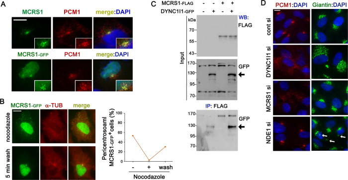
Figure 1. MCRS1 is required for proper distribution of centriolar satellites.
(A) Immunofluorescence images showing the distribution of endogenous MCRS1 and the centriolar satellite marker PCM1 (upper panels). RPE1 cells transiently expressing EGFP-tagged MCRS1 were stained with PCM1 antibody (lower panels). (B) RPE1 cells transiently expressing EGFP-tagged MCRS1 were treated with nocodazole for 1 hr, and then stained with α-Tubulin antibody with or without nocodazole washout (5 min wash). The graph shows quantification of cells exhibiting pericentrosomal accumulation of MCRS1-GFP-positive granules: (-) without nocodazole treatment, (+) after 1 hr nocodazole treatment, and (wash) after 1 hr nocodazole treatment followed by 5 min washout. (C) HEK293T cells were transfected with the indicated plasmids for 16 hr, and then cell lysates were immunoprecipitated with anti-FLAG antibody conjugated with agarose beads. The resulting precipitates and input lysates were immunoblotted with the indicated antibodies. Arrow indicates bands at the molecular weight of DYNC1l1-GFP. (D) Immunofluorescence images showing the distribution of PCM1-containing granules and the Golgi complex after transfection of the indicated siRNAs for 3 days. Scale bars represent 10 μm.

