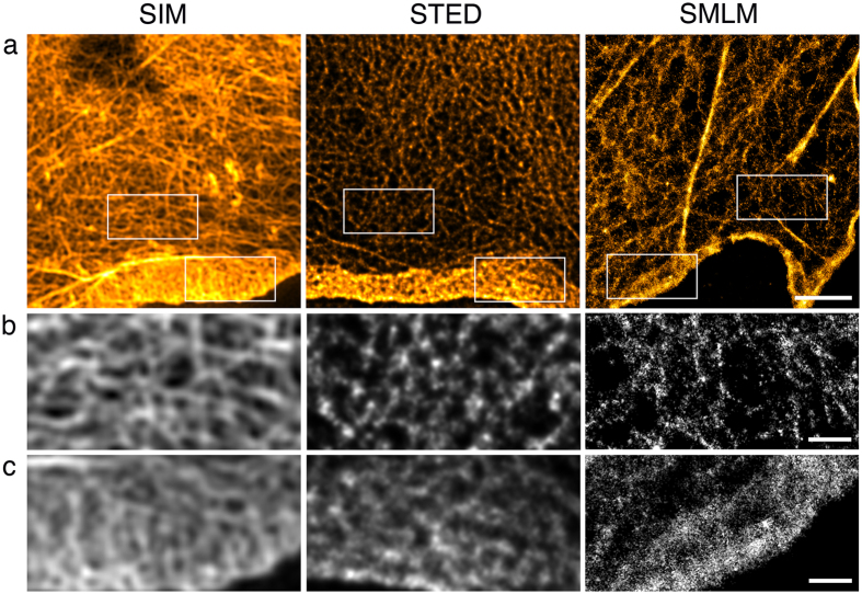
Figure 3. Actin in COS7 cells.
Actin was detected with phalloidin coupled to Alexa Fluor 488 for SIM and STED and Alexa Fluor 647 for SMLM. 100% available depletion laser power was used for STED. (a) Single optical section through the cell periphery. Boxed areas depict parts of the lamella (b) and the lamellipodium (c). The STED image has been 2D deconvolved. Scale bar, 2 μm. (b) Fine structure of the lamella. Scale bar, 0.5 μm. (c) Fine structure of the lamellipodium. Scale bar, 0.5 μm.

