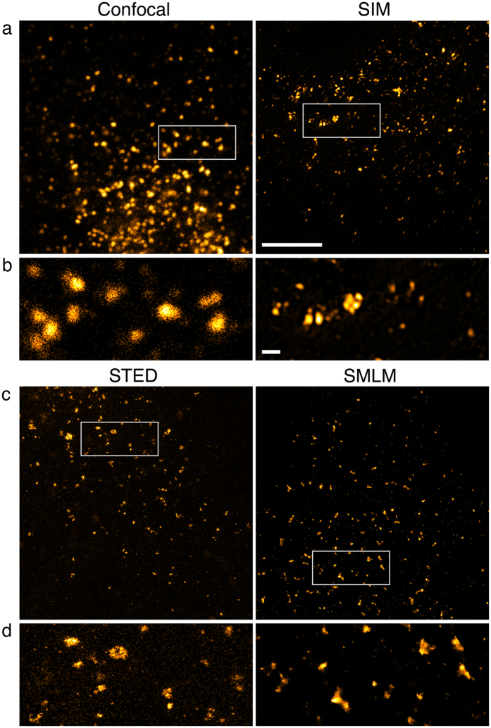
Figure 4. Transport vesicles in COS7 cells.
Sec31A was detected with primary and secondary antibodies, the latter coupled to Alexa Fluor 488 for confocal imaging, SIM and STED and Alexa Fluor 647 for SMLM. 100% available depletion laser power was used for STED. (a,c) Overview of vesicles close to the nucleus in a single optical section. Boxed areas are magnified in (b,d). Scale bar, 5 μm. (b,d) Vesicles of various sizes with a little out of focus background visible as weak, small foci in STED. Scale bar, 0.5 μm.

