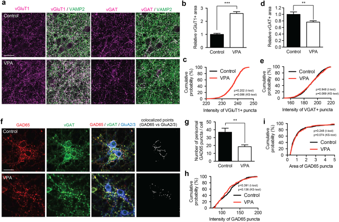
Figure 4. Differential modification of glutamatergic and GABAergic presynaptic terminals in response to VPA in cultured neurons.
(a) Representative images of vGluT1/VAMP2 or vGAT/VAMP2 protein immunocytochemistry in control or in vitro VPA model. The density of vGluT1 positive presynapses was increased and density of vGAT positive presynapses decreased. Scale bar = 20 μm. (b–e) Quantification of vGluT1- and vGAT-positive areas and intensity distribution of punta in control and VPA-treated cultures (≥24 images from 3 independent experiments). (f) Representative images of GAD65 and vGAT protein immunocytochemistry in control or in vitro VPA model (anti-GluA2/3 immunereactivity was used to outline neuronal cell somata). (g–i) The number of GAD65 positive perisomatic puncta per cell was reduced in VPA treated cultures, whereas intensity and size of GAD65 puncta were not altered. Scale bar =20 μm (≥15 images from 3 independent experiments).

