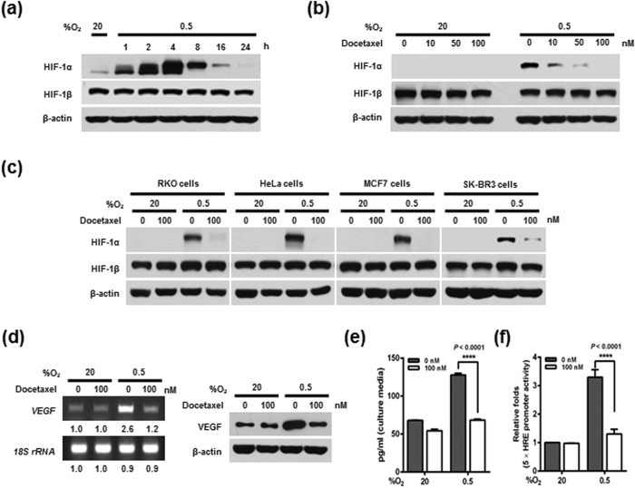
Figure 1. Effects of docetaxel on HIF-1α expression and transcriptional activation in cancer cells under hypoxia.
(a) MDA-MB-231 cells were exposed to 0.5% O2 for 24 h and harvested at the indicated times. Whole-cell lysates were analyzed by immunoblotting for the indicated proteins. (b) MDA-MB-231 cells were treated with different concentrations of docetaxel (0–100 nM) for 16 h and exposed to 20% or 0.5% O2. After a 4-h incubation, cells were harvested, and cell lysates were analyzed by immunoblotting for the indicated proteins. (c) RKO, HeLa, MCF7, and SK-BR3 cells were incubated with or without 100 nM docetaxel for 16 h and exposed to 20% or 0.5% O2. After a 4-h incubation, cells were harvested, and whole-cell lysates were analyzed by immunoblotting for the indicated proteins. (d) MDA-MB-231 cells were incubated with or without 100 nM docetaxel for 16 h, exposed to 20% or 0.5% O2 for 24 h, and then harvested. RT-PCR (left panel) was used to amplify VEGF mRNA and 18S rRNA, and immunoblot analysis (right panel) was used to detect VEGF and β-actin proteins. Band intensities of RT-PCR products from cells cultured under hypoxic conditions relative to those from cells in normoxic conditions were quantified using Image J (NIH). (e) MDA-MB-231 cells were incubated with or without 100 nM docetaxel for 16 h and exposed to 20% or 0.5% O2. After a 24-h incubation, conditioned media were harvested. Secreted VEGF in conditioned media was analyzed by ELISA. Data are presented as means ± SD (****P < 0.0001; ANOVA). (f) MDA-MB-231 cells co-transfected with p5 × HRE-luc and pCMV-β-galactosidase were cultured for 16 h, then incubated with or without 100 nM docetaxel for 16 h, and exposed to 20% or 0.5% O2 for 4 h. Luciferase activity was normalized to that of β-galactosidase. Data are presented as means ± SD (****P < 0.0001; ANOVA).

