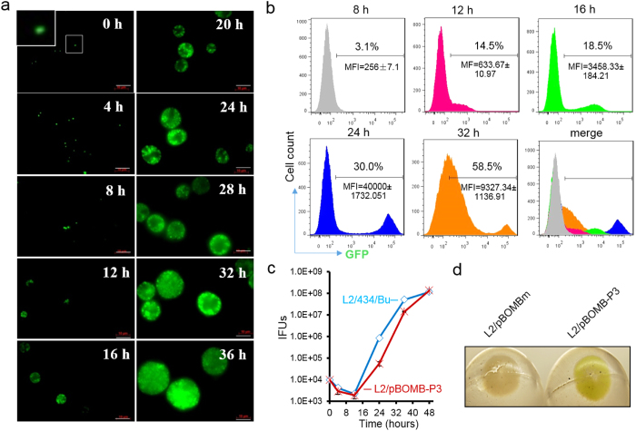
Figure 6. Inspecting P3-GFP levels during the course of the C. trachomatis infection.
(a) Time course of GFP expression in HeLa cells visualized by fluorescent microscopy. L2/pBOMB-P3 infected HeLa cells were imaged at the times indicated. The inset highlights GFP-expressing C. trachomatis organisms. Bar = 10 μM. (b) Flow cytometric data corroborate microscopic images in (a), showing changes in GFP expression in C. trachomatis infected HeLa cells. GFP-expressing cell population is indicated as percentage. The average mean fluorescence intensity (MFI) and the standard deviation at each time is shown. (c) One-step growth curve of C. trachomatis strains L2/434/Bu and L2/pBOMB-P3. (d) Isolated L2/pBOMB-P3 organisms, but not L2/pBOMBm, appear to be green-colored by direct observation.

