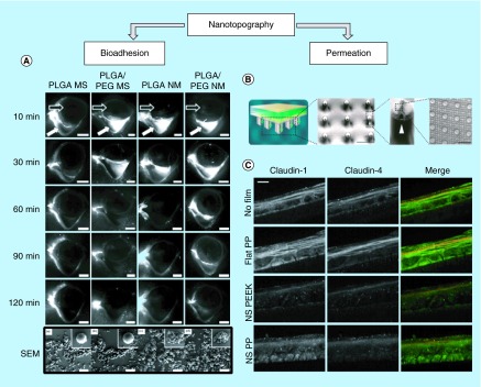
Figure 2. . Nanotopography-inspired approaches to enhance bioadhesion and permeation.
(A) Fluorescence images of the preocular surface of the rabbit eye after administration of particles tagged with Nile Red and scanning electron microscope (SEM) images of particles. Compared with bare PLGA or PLGA/PEG microspheres, microparticles with nanotopography achieved greater preocular retention time in vivo. White arrow indicates lower fornix of the rabbit eye and black arrow indicates the location of the eyeball. The bottom row shows SEM images of the particles. Scale bar = 5 mm for the top five rows and 20 µm for bottom row of SEM images [27]. (B) Nanostructured microneedles were fabricated by draping polymeric nanostructured film on a microneedle array. Scale bars = 300 µm and 3 µm in order [28]. (C) Nanostructures made with polypropylene or polyether ether ketone downregulate expression of tight junction proteins, such as Claudin-1 and 4, in cultured human keratinocytes as demonstrated by immunohistochemical staining. Scale bar = 10 µm [28].
PEG: Polyethylene glycol; PLGA: Poly(lactic-co-glycolic acid); PP: Polypropylene; NS: Nanostructured, PEEK: Polyether ether ketone.
Adapted with permission from [27,28] © Elsevier (2014) and © American Chemical Society (2015).

