Fig. 6.
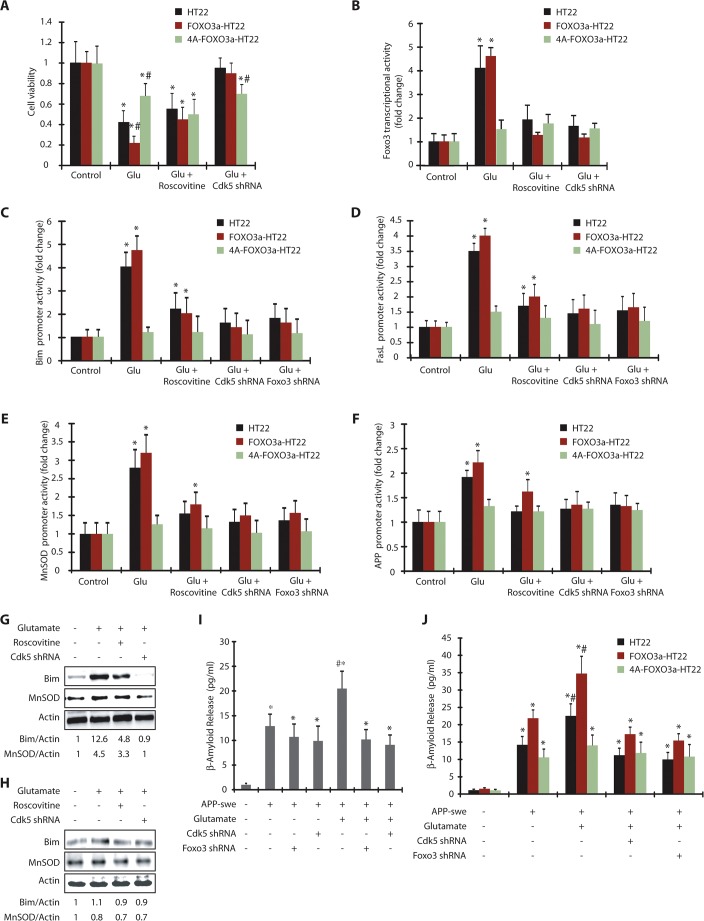
Cdk5-mediated activation of Foxo3 and its downstream genes are phosphorylation-dependent. (A) HT22, FOXO3a-HT22 and 4A-FOXO3a-HT22 cells were treated with glutamate, roscovitine and Cdk5 shRNA and cell viability tested by using an MTT assay. *P<0.01, compared with untreated HT22 cells; #P<0.01, compared with glutamate-treated HT22 cells (Student's t-test). (B) HT22, FOXO3a-HT22 and 4A-FOXO3a-HT22 cells transfected with pGL3-based Foxo3 promoter plasmid were treated with glutamate in the presence or absence of roscovitine or Cdk5 shRNA and the promoter activity measured. *P<0.01, compared with untreated HT22 cells (Student's t-test). (C) Bim promoter activity was measured in glutamate, roscovitine, Foxo3 shRNA and Cdk5 shRNA-treated HT22, FOXO3a-HT22 and 4A-FOXO3a-HT22 cells. *P<0.01, compared with untreated HT22 cells. (D) FasL promoter activity was measured in glutamate, roscovitine, Foxo3-shRNA- and Cdk5-shRNA-treated HT22, FOXO3a-HT22 and 4A-FOXO3a-HT22 cells. *P<0.01, compared with untreated HT22 cells (Student's t-test). (E) HT22, FOXO3a-HT22 and 4A-FOXO3a-HT22 cells were transfected with pGL3-based MnSOD promoter, followed by various treatments and the promoter activity measured. *P<0.01, compared with untreated HT22 cells (Student's t-test). (F) APP promoter activity was measured in glutamate, roscovitine, Foxo3 shRNA and Cdk5-shRNA-treated cells *P<0.01, compared with untreated cells (Student's t-test). (G) FOXO3a-HT22 cells pretreated with roscovitine or Cdk5 shRNA were treated with glutamate for 18 h. The total levels of Bim or MnSOD were analyzed. (H) 4A-FOXO3a-HT22 cells pretreated with roscovitine or Cdk5 shRNA were treated with glutamate (18 h). The total levels of Bim or MnSOD were analyzed. (I) APP-swe plasmid was transfected into HT22 cells for 40 h, followed by 18 h of glutamate treatment. Cdk5 shRNA or Foxo3 shRNA were added 30 h prior to glutamate treatment. β-amyloid (1-42) released into the medium was measured as described in the Materials and Methods. *P<0.01, compared with untreated HT22 cells; #P<0.01, compared with glutamate-treated HT22 cells (Student's t-test). (J) APP-swe was transfected into HT22, FOXO3a-HT22 and 4A-FOXO3a-HT22 cells, followed by various treatments. β-amyloid(1–42) released into the medium was measured. Graphical results are mean±s.e.m. Each experiment was repeated at least three independent times.

