FIGURE 4.
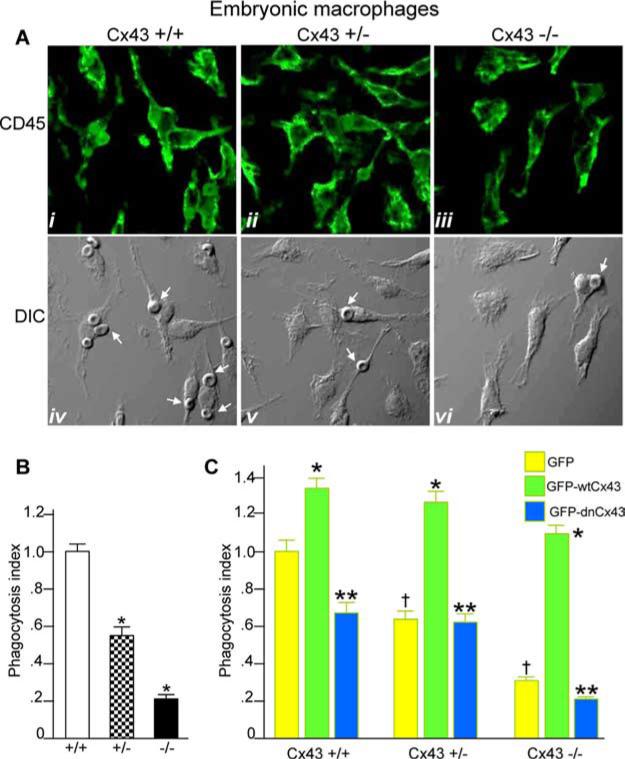
Expression of adenovirus GFP-Cx43 reverses the impairment in phagocytosis observed in Cx43-deficient macrophages. A, Macrophages were harvested from the livers of embryonic Cx43+/+, Cx43+/−, and Cx43−/− mice at gestational age embryonic days 16–18 as described in Materials and Methods, immunostained with Abs against the macrophage marker CD45, and examined by confocal microscopy. Representative CD45 staining (i-iii) and the corresponding DIC images (iv-vi) are shown. B, Embryonic macrophages were harvested from livers of Cx43+/+, Cx43+/−, and Cx43−/− mice and allowed to internalize opsonized RBCs as described in Materials and Methods. The phagocytosis index (no. of cells with at least one internalized RBC per 100 cells relative to wild-type macrophages) is shown. Representative of five separate experiments. *, p < 0.05 vs wild-type macrophages. C, Macrophages were harvested from Cx43+/+, Cx43+/−, and Cx43−/− mice and infected with adenoviruses expressing GFP (yellow bars), GFP-wtCx43 (green bars), or GFP-dnCx43 (blue bars). Cells were then allowed to undergo phagocytosis of opsonized RBCs as described in Materials and Methods. The phagocytosis index (number of macrophages with at least one internalized particle per 100 cells relative to GFP-infected macrophages from each strain) is shown. †, p < 0.05 vs GFP-infected cells from wild-type mice. Note that in each case the rate of phagocytosis was decreased in GFP-infected cells from +/− and −/− mice vs +/+ mice. *, p < 0.05 vs GFP-infected macrophages for each strain. Note that in each case infection with GFP-wtCx43 leads to a significant increase in phagocytosis; **, p < 0.05 vs GFP-wtCx43. Note that for each mouse strain, infection with GFP-dnCx43 leads to a significant decrease in the rate of phagocytosis compared with infection with wild-type Cx43. Representative of at least five separate experiments with more than five mice and 100 cells per group.

